Biomechanical Diagnostic
Pre-Planning and Outcome Tools to improve Musculoskeletal Surgery
Within the BioMechTools program we will develop diagnostic and evaluation tools to quantify the degenerative status of orthopaedic patients. Our focus lies on the lower extremities. This project will open new research fields related to biomechanical patient assessment and modelling of musculoskeletal pathologies.
Project Description
The aetiology of many musculoskeletal diseases is related to biomechanical factors. However, the tools used by clinicians and researchers to assess the biomechanical condition of patients are often crude and subjective, leading to non-optimal patient analyses and care. For example, clinicians usually base their diagnosis on their own, subjective, interpretation of static imaging data like MRI-images and CT-scans. Yet, it is much more powerful to visualize the pathological tissue in a dynamic manner, i.e. a tear in a meniscus is easier to see when the meniscus is moving than when it is imaged statically.
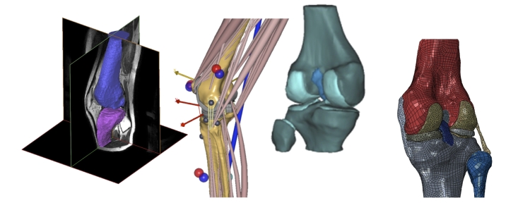
The Orthopaedic Research Laboratory, in collaboration with the Laboratory of Biomechanical Engineering (University of Twente), has been very successful in delivering imaging modalities and in generating patient specific musculoskeletal and finite element models. These types of models can be used to analyse the pathology in detail and to assess the biomechanical consequences of various surgical scenarios. See, for example, our research line ‘Musculoskeletal Biomechanics‘ and the ‘TLEMsafe‘ project.
With the BioMechTools project we aim to develop MRI and Ultrasound tools to quantify tissue strains under dynamic circumstances. Innovations related to imaging, sensor technology and biomechanical modelling will be utilized to generate versatile, accurate and objective methods to quantify the (pathological) musculoskeletal condition of the lower extremity of patients in a unique manner. These advanced diagnostic, pre-planning and outcome tools will provide clinicians and researchers with detailed biomechanical analysis about abnormal tissue deformations, pathological loading of the joints, abnormal stresses in the hard and soft tissues, and aberrant joint kinematics.
Our research will be done in collaboration with the of the Technical University of Eindhoven and the Radiology department of the Academic Medical Centre Amsterdam for the dynamic MRI imaging and with the ‘Medical UltraSound Imaging Center‘ of the Radboudumc for the ultrasound techniques.
Our key objectives are:
- develop and validate image based 3-D volumetric elastographic diagnostic methods that can quantify normal and pathological conditions of tissues under dynamic loading and which can be linked to biomechanical modelling tools
- create an ultrasound-based system to assess internal joint kinematics which can be used as a diagnostic tool for clinicians and researchers and which is a validation tool for biomechanical modelling
- generate and validate an ambulant functional (force and kinematic) diagnostic system which is easy to use and which can be used to provide input data for biomechanical models
- create and validate a new modelling approach that integrates muscle-models with finite element models at a highly personalized level
- generate biomechanical models which have personalized mechanical properties of the hard and soft tissues
- demonstrate the applicability of the personalized diagnostic and pre-planning platform by application to healthy individuals and to patients
Principle Investigator:
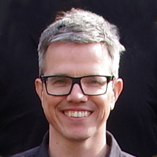
Nico Verdonschot, Prof. Dr. Ir.

Co-Supervisor:
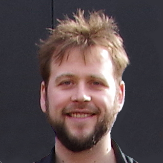
André Sprengers, Dr.
Co-Supervisor:
Jasper Homminga, Dr. Ir.

Co-Supervisor:
Dennis Janssen, Dr.